 | |||
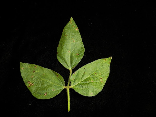
| Figure 1. Soybean plant exhibiting symptoms of Phyllosticta leaf spot and iron deficiency chlorosis. Photo: Angie Peltier |
A mystery disease in northwest and west-central Minnesota soybeans. A couple of weeks ago now, unifoliate and the first trifoliate soybean leaves in research fields in Crookston and Barrett, MN (note: different varieties, different maturities) began exhibiting relatively uniform rounded brownish lesions. While not every plant had symptoms, the relative uniformity in symptoms across the field and small, brownish lesions led one to suspect an abiotic (not caused by a living organism) cause.
Fast forward a couple of weeks, and symptoms are no longer confined to unifoliate leaves or the first trifoliate leaf, but several trifoliate leaves are now showing symptoms (Figure 1). Specifically, tan lesions that are bordered by larger leaf veins and ringed in a dark brown border.
While the symptoms can easily be confused with frogeye leaf spot lesions (Figure 2), young frogeye lesions tend to be more round and are bordered by a more purplish-brown border. In addition, although soybean is susceptible to the pathogen that causes frogeye leaf spot at any growth stage, symptoms tend to appear later in the growing season when there are warmer temperatures and relative humidity is higher.
 |
| Figure 2. Frogeye leaf spot lesions on a soybean leaf. Notice how the lesions are very round and are ringed in purplish-brown. Photo: Angie Peltier |
One sure-fire way to tell the two diseases apart is to place leaves in a moist chamber and see what grows. In my case, I collected multiple symptomatic leaves, placing half in each of two 'ziplock'-style plastic bags. Inside the bag, but not touching the leaves, was also added a wet paper towel. After inflating and then sealing the bags, one was placed on a counter at room temperature and one in a refrigerator at approximately 35 degrees.
In fewer than 24 hours I had my answer when small, round, black fungal fruiting structures called pycnidia formed on the upper leaf surface on some of the lesions (Figure 3). Pycnidia hold asexual spores of the fungus that causes Phyllosticta leaf spot.
 |
| Figure 3. Phyllosticta leaf spot lesion with pycnidia (red arrow). Seeing pycnidia is one way to determine that the lesions you are observing aren't frogeye leaf spot. Photo: Angie Peltier |
Had there been a greyish, fuzzy fungal growth that developed inside of the lesions, Cercospora sojina, the fungus that causes frogeye leaf spot was more likely to be the cause of the lesions (Figure 4).
 |
| Figure 4. Sporulation of the frogeye leaf spot pathogen, Cercospora sojina (blue arrow). Photo: Daren Mueller, Iowa State University, Bugwood.org |
One way to know what is causing disease symptoms in any plant is to send a sample to the UMN Plant Disease Clinic for examination and testing by diagnosticians.
Disease management.
The fungus that causes Phyllosticta leaf spot (Phyllosticta sojicola) is thought to survive in both infested residue and in seed that had been previously infected. It is not known whether spring 2023 was simply more conducive for disease development, the varieties planted were more susceptible than others, or whether the seed originated from an area with a lot of Phyllosticta leaf spot and therefore carried primary inoculum from a 2022 infection.
Regardless, cool, moist conditions are thought to favor disease development, and in years in which symptoms are observed, yield loss has been rare. As Phyllosticta leaf spot is a polycyclic disease (a disease with multiple cycles of infection throughout the growing season), if disease symptoms continue to spread, I will put out a small fungicide trial at the UMN Northwest Research & Outreach Center on plants not yet dedicated to an unrelated research project.
References.
Hartman, G.L., Rupe, J.C., Sikora, E.J., Domier, L.L., Davis, J.A. and Steffey, K. L. eds. 2015. Compendium of Soybean Diseases and Pests. Fifth ed. APS Press. St. Paul, MN.
Comments
Post a Comment